


|
|
|
|
|
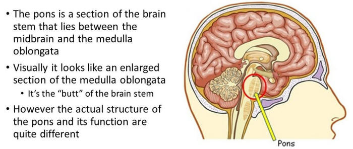
An important aspect in the explanation of dreaming is an understanding of the portions of the brain, which work together to create and terminate dreams and the dreaming state (i.e. REM sleep). A variety of nuclei and brain regions are involved in the initial production of the REM state, including the amygdala, hippocampus, right temporal lobe, and especially the brainstem nuclei located in the lateral and medial pons. REM sleep begins with signals from the base of the brain called the pons. The pons are associated with retitcular activity, and send signals to the region of the brain called the thalamus, which intern passes them along to the cerebral cortex for interpretation. While the pons are sending signal to the thalamus, they are also sending signals to the spinal chord, which shut off motor neurons making it impossible for the body to move during sleep and dreaming. (reference)
Similarly, cerebral blood flow increases in the right temporal and parietal regions during REM sleep. These stages are followed by abnormal and enhanced activity in the right temporal and temporal-occippital area, which causes an increase in dreaming and REM sleep for an uncharacteristically long period of time. The brainstem is the most important actor in dream production, for it controls the steady and specific oscillations between REM and non-REM or NREM sleep, the same system that controls body and mind activation in waking periods. Overall, dreaming and REM sleep are associated with high levels of activity in the brainstem, occipital lobe, and other nuclei.
Though these parts of the brain are all active during REM sleep, it is important to know that the two hemispheres of the brain work unilaterally in dreaming. The right hemisphere of the brain actually creates and displays the dream, shown by an increase in blood flow and electrophysiological stimulation in that hemisphere during REM. The right hemisphere uses a form of visual-spatial and emotional language, which creates the themes and images of the dream through remembered emotions. These sorts of memories, such as those used as the material for dreaming, and those that later will become the remembered dream are called lateralized memories, for they are only remembered by one hemisphere of the brain, the right.
Therefore, due to the lateralized remembrance of dreams, the left hemisphere of the brain must access the memories of the right brain when a person is asked to describe a dream or even to remember it. To access the memory banks of the right hemisphere, the left hemisphere must use the corpus callosum or anterior commissure. The left hemisphere of the brain is involved primarily in the encoding and recall of verbal, temporal sequential, and language related memories. As a result, dream interpretation becomes very difficult due to the fact that the left hemisphere is forced to interpret what the right brain has created using a language the left brain does not understand. This accounts for the often sporadic and non-sequential order of dreams, for the right brain, which created the dream, cannot establish sequences. Therefore, the left hemisphere must try to put a dream in order when it is interpreting it. On rare occasions, a person may experience a dream during non-REM sleep, a dream created entirely by the left hemisphere of the brain. These dreams are typically devoid of imagery, being more like a running monologue and interestingly, it is during this type of dreaming that one may talk in his/her sleep.
A great variety of neurotransmitters are involved in driving wakefulness and sleep, including histamine, dopamine, norepinephrine, serotonin, glutamate, orexin and acetylcholine, among others. While none of these neurotransmission processes is individually necessary, they all appear to contribute in some way. Histamine in particular is sometimes referred to as the master wakefulness-promoting neurotransmitter, exhibiting high activity during wakefulness, decreasing activity during NREM sleep, and its lowest levels during REM sleep. That is why histamine-blocking antihistamine medications cause drowsiness and increase NREM sleep. Serotonin activity promotes wakefulness, increases sleep-onset latency or the length of time it takes to fall asleep and decreases REM sleep. Acetylcholine activity in the reticular activating system of the brainstem stimulates activity in the forebrain and cerebral cortex, encouraging alertness and wakefulness, although it also appears to be active during REM sleep. Dopamine activity sometimes seems to promote wakefulness and sometimes sleep, so its role is still far from clear. (reference)
We are at least as dream deprived as we are sleep deprived. Many of the health concerns attributed to sleep loss result from a silent epidemic of REM sleep deprivation. REM/dream loss is an unrecognized public health hazard that silently wreaks havoc with our lives, contributing to illness, depression, and an erosion of consciousness. Emerging evidence suggests that REM/dreaming affects immune function, memory consolidation, mood regulation, as well as transpersonal, religious, or spiritual experiences. (reference)
See also: Bibliography, Booze and dreams, Dream cycles, Dream glossary, Dream hacking, Dream recall, Dreams and brain disorders, Dreams as a source of inspiration, Essential oils, Food and dreams, Herbs for dreaming, Hypnagogic state, Lucid dreaming, Neuroprotective agents, Precognitive dreams, Psychic dreams, Recurring dreams, Shamanic dreaming, Sleeping brain, Sleep deprivation, Weed and dreams, WILD, Yoga Nidra